
Giardia and Cryptosporidium Detection: Filtration Enrichment Methods for Laboratories
Why Filtration Enrichment Is Critical for Giardia and Cryptosporidium Detection
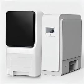
Giardia and Cryptosporidium (oo)cysts are typically found in surface water, groundwater under the influence of surface water, and even treated drinking water at very low concentrations. A single liter of water may contain only a few cysts or oocysts. Without concentration, the probability of missing them entirely during analysis is unacceptably high. Filtration enrichment allows a laboratory to process 10 liters, 50 liters, or more of water, capturing and retaining the parasites on a filter while letting the water pass through. The retained material is then eluted and further concentrated, resulting in a small final volume that can be examined in its entirety. The US EPA Method 1623 and similar procedures in Standard Methods for the Examination of Water and Wastewater rely on this principle to achieve the sensitivity required for regulatory monitoring and outbreak investigations.
Common Filtration Enrichment Methods in the Laboratory
No single filtration approach works best for all sample types. Laboratories typically choose among cartridge filtration, membrane filtration, and flocculation-based concentration, depending on sample volume, turbidity, and available equipment. The table below compares the three most widely used Giardia and Cryptosporidium detection system enrichment techniques.
| Method | Principle | Typical Volume Processed | Recovery Efficiency | Strengths | Limitations |
|---|---|---|---|---|---|
| Cartridge Filtration | Depth filtration through a pleated, high-surface-area cartridge (1 µm nominal pore size); retained organisms are eluted by backwashing and washing the cartridge. | 10–1,000 L | 30–70%, highly variable with water quality | Handles large volumes, suitable for very low contamination levels, field-deployable | Labor-intensive elution, recovery drops with turbidity, cartridge cost |
| Membrane Filtration | Direct filtration through a flat disc membrane (1–3 µm pore size); organisms are trapped on the surface, then eluted by scraping or sonication. | 1–20 L | 40–80% for clean water, decreases with turbidity | Simple setup, good recovery in low-turbidity samples, lower cost per sample | Limited volume, clogging with particulates, manual membrane handling |
| Calcium Carbonate Flocculation | Chemical flocculation using calcium carbonate to co-precipitate cysts and oocysts; flocs are settled and then dissolved for concentration. | 10–100 L | 50–80%, consistent across turbidity levels | Effective for turbid water, less equipment dependence, can process large volumes | Requires chemical handling, multiple centrifugation steps, longer procedure |
According to the Standard Methods for the Examination of Water and Wastewater, the choice of concentration technique should be validated for the specific water matrix to ensure reliable recovery and consistent detection limits.
Step-by-Step Water Testing Workflow Using Filtration Enrichment
While details vary with the chosen method, the general workflow for a Giardia and Cryptosporidium detection system includes the following steps:
- Sample Collection and Preservation: Collect the water sample following aseptic technique. Large-volume samples are often preserved with sodium thiosulfate (if chlorinated) and kept cool during transport.
- Filtration/Concentration: Pass the water through the selected filter or perform flocculation. For cartridge filters, run water through until the desired volume is reached or the flow rate drops significantly. For membrane filters, vacuum filtration is common.
- Elution and Washing: Recover captured organisms from the filter matrix. Cartridge elution involves mechanical agitation and backwashing with an eluting solution; membrane filters are typically scraped or vortexed in a small volume of buffer.
- Secondary Concentration (if needed): Further reduce the eluate volume by centrifugation or additional membrane filtration to achieve a final volume suitable for immunomagnetic separation (IMS) or direct examination.
- Immunomagnetic Separation (IMS): Use antibody-coated magnetic beads to specifically capture Giardia cysts and Cryptosporidium oocysts from the concentrate, removing background debris.
- Staining and Microscopy: Apply fluorescently labeled monoclonal antibodies (e.g., FITC-conjugated) and DAPI for nuclear staining. Examine by epifluorescence and differential interference contrast microscopy.
- Results Interpretation: Identify and enumerate (oo)cysts based on size, shape, fluorescence pattern, and internal morphological features as described in regulatory methods.
Automated systems that integrate filtration, elution, and IMS are available and can reduce hands-on time while improving reproducibility. Even with automation, careful adherence to quality control protocols is essential.
Lab Safety Guidelines for Giardia and Cryptosporidium Testing
Giardia and Cryptosporidium are zoonotic pathogens, and Cryptosporidium oocysts in particular are resistant to many common disinfectants. Laboratories must handle all water concentrates as potentially infectious. Basic safety practices include:
- Work inside a certified Class II biological safety cabinet when handling concentrated samples, especially during elution, centrifugation, and IMS steps.
- Wear appropriate personal protective equipment: laboratory coat, gloves, and eye protection. Use a face shield if there is a splash risk.
- Avoid creating aerosols during vortexing, centrifugation, or filter scraping. Use sealed rotors and safety cups for centrifuges.
- Decontaminate work surfaces with a suitable disinfectant effective against Cryptosporidium, such as 6% hydrogen peroxide or a 1:10 dilution of household bleach (freshly prepared) with adequate contact time.
- Autoclave all disposable materials that have contacted sample concentrates before disposal.
- Maintain strict separation between pre‑enrichment and post‑enrichment work areas to prevent cross-contamination.
All personnel should receive specific training on the risks associated with Cryptosporidium and the heightened need for disinfection vigilance.
Interpreting Results and Reporting Needs
Interpreting Giardia and Cryptosporidium detection system results requires more than a positive/negative call. Regulatory reporting under the Long Term 2 Enhanced Surface Water Treatment Rule (LT2) or similar frameworks in other countries demands quantitative data and method quality control information.
Key reporting elements include:
- Number of confirmed (oo)cysts counted in the entire concentrate, broken down by organism.
- Sample volume processed and the equivalent volume examined (after all concentration steps).
- Recovery efficiency from positive control samples (method‐spike controls) processed alongside the field samples.
- Method detection limit (MDL) calculated based on recovery and volume.
- Any procedural deviations, matrix interferences, or unusual observations.
Laboratories must calculate the concentration in organisms per liter using the recovery efficiency as a correction factor. For example: Concentration = (number of (oo)cysts counted) / (volume examined × recovery efficiency). A clear, standardized reporting format that includes all these elements ensures that water utilities, public health authorities, and regulators can properly interpret the data.
How to Select a Giardia and Cryptosporidium Detection System
When evaluating a filtration enrichment system or integrated detection platform, use the following checklist to compare options against your laboratory’s operational needs:
- Sample Volume Capacity: Can the system handle the required sample volumes (e.g., 10 L, 50 L, 100 L) within a practical time frame?
- Compatibility with Water Types: Does the method work with source water, finished drinking water, turbid/recreational water, and wastewater? Check the manufacturer’s validation for each matrix.
- Recovery Efficiency and Consistency: Review published performance data for Giardia and Cryptosporidium in relevant water types. Look for recovery of at least 30–50% with low variability.
- Labor and Workflow Integration: How much hands-on time is needed? Can the system be left unattended for filtration? Is the elution process manual or automated?
- Safety Features: Enclosed systems that minimize aerosol generation and direct contact with concentrates reduce laboratory‑acquired infection risk.
- Sample Throughput: Number of samples that can be processed per day. Consider peak periods (e.g., after heavy rainfall) when demand spikes.
- Cost per Sample: Include consumables (filters, cartridges, reagents, IMS beads), maintenance, and labor.
- Regulatory Acceptance: Ensure the method is accepted by the relevant regulatory authority (e.g., EPA approval for compliance monitoring) if results will be used for legal purposes.
- Technical Support and Training: Availability of application support, initial training, and troubleshooting assistance.
A well-chosen Giardia and Cryptosporidium detection system streamlines the entire workflow from sample collection to report generation, while minimizing reproducibility issues and safety risks.
Final Considerations
Filtration enrichment remains the cornerstone of reliable Giardia and Cryptosporidium detection in water. The right method and system selection depend on your typical sample volumes, water quality, and regulatory obligations. By following a validated workflow and maintaining rigorous safety practices, laboratories can provide accurate data that supports public health decisions and water quality management. As detection technology evolves, automated and integrated platforms continue to reduce hands-on time and improve measurement precision, making routine monitoring more feasible for a wider range of facilities.
For system-level planning, our Laboratory Equipment Solution page can help buyers connect equipment selection with real hospital or laboratory workflows. Related equipment pages include Giardia and Cryptosporidium Filtration Enrichment and Detection System and HD-F3P Three-Place Microbial Filtration Detection System.